Anatomy and Function of the Meniscus

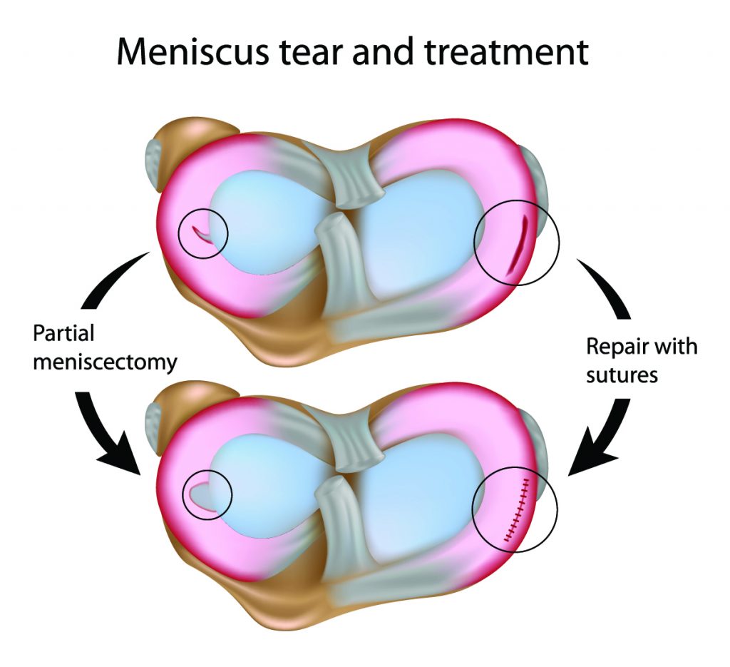
The menisci are C-shaped pieces of fibrocartilage that act as shock absorbers and stabilizers within the knee joint. They are located between the femur (thigh bone) and the tibia (shin bone), playing a crucial role in maintaining joint health and function.
Location and Structure
The menisci are situated on the tibial plateau, the top surface of the tibia. There are two menisci in each knee: the medial meniscus, located on the inner side of the knee, and the lateral meniscus, situated on the outer side. Both menisci are wedge-shaped, with a thicker outer edge and a thinner inner edge. They are primarily composed of type I collagen fibers, arranged in a circumferential pattern, which provides strength and resilience. The menisci are avascular, meaning they lack a direct blood supply, except for a small peripheral zone.
Functions of the Menisci
- Load Distribution: The menisci act as shock absorbers, distributing weight evenly across the knee joint during activities like walking, running, and jumping. By spreading the load, they protect the articular cartilage, which lines the joint surfaces, from excessive wear and tear.
- Joint Stability: The menisci contribute to the stability of the knee joint by deepening the articulation between the femur and tibia. This increased congruency helps to prevent the femur from sliding off the tibia during movement.
- Shock Absorption: The menisci act as cushions, absorbing shock and reducing the impact forces transmitted to the knee joint during activities. This cushioning effect helps to protect the joint from injury.
Blood Supply to the Meniscus
The menisci receive their blood supply from a network of small vessels that originate from the surrounding joint capsule. However, the blood supply is limited and varies depending on the location within the meniscus. The meniscus is divided into three zones based on its blood supply:
- Red Zone: This zone is located at the outer edge of the meniscus and receives a rich blood supply from the surrounding capsule. This zone has the best potential for healing after injury.
- Red-White Zone: This zone is located in the middle of the meniscus and receives a limited blood supply. This zone has a moderate potential for healing.
- White Zone: This zone is located in the inner portion of the meniscus and lacks a direct blood supply. This zone has a poor potential for healing.
Types and Causes of Meniscus Tears

Meniscus tears are common injuries, especially among athletes and individuals engaging in physically demanding activities. Understanding the different types of meniscus tears and their causes is crucial for proper diagnosis and treatment.
Types of Meniscus Tears
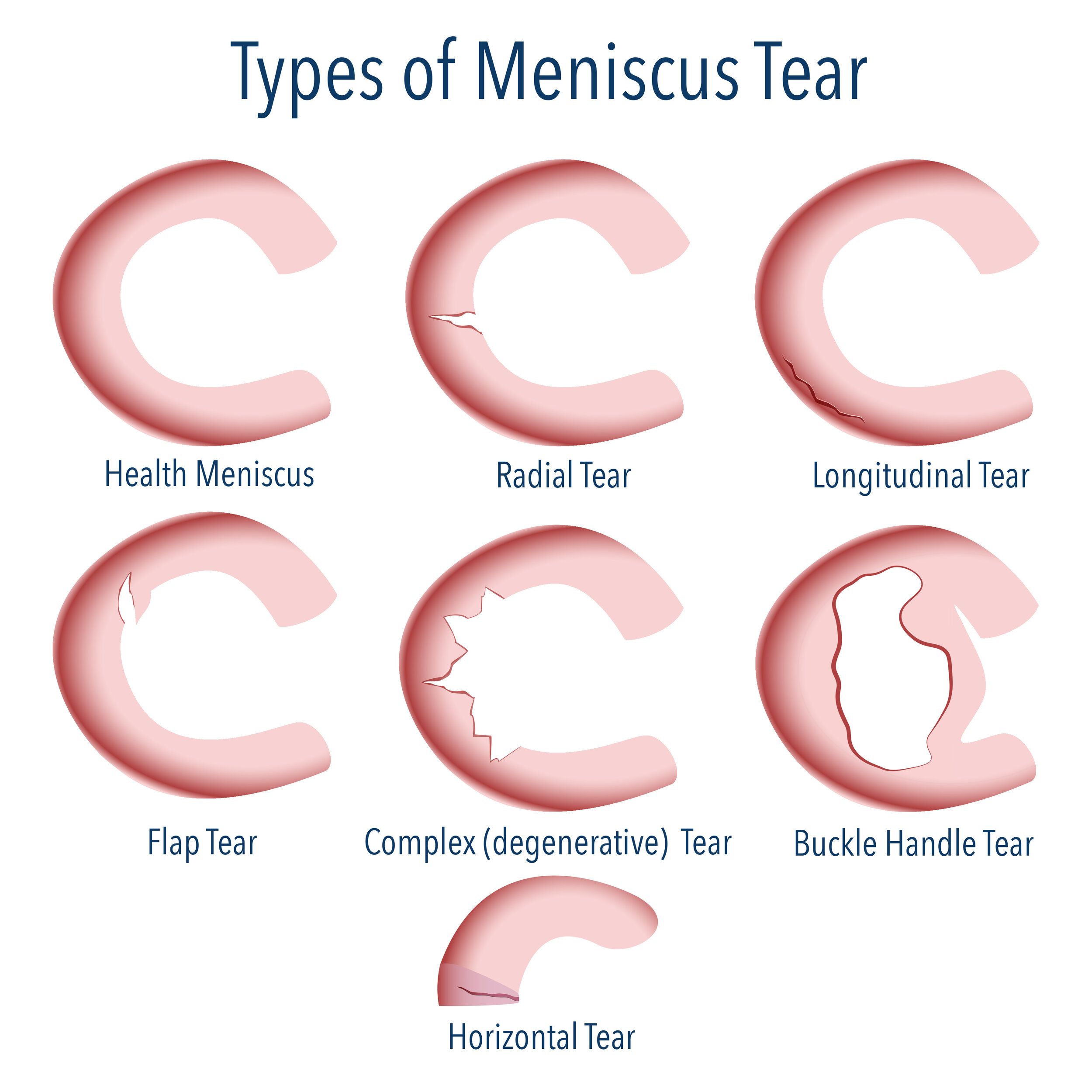
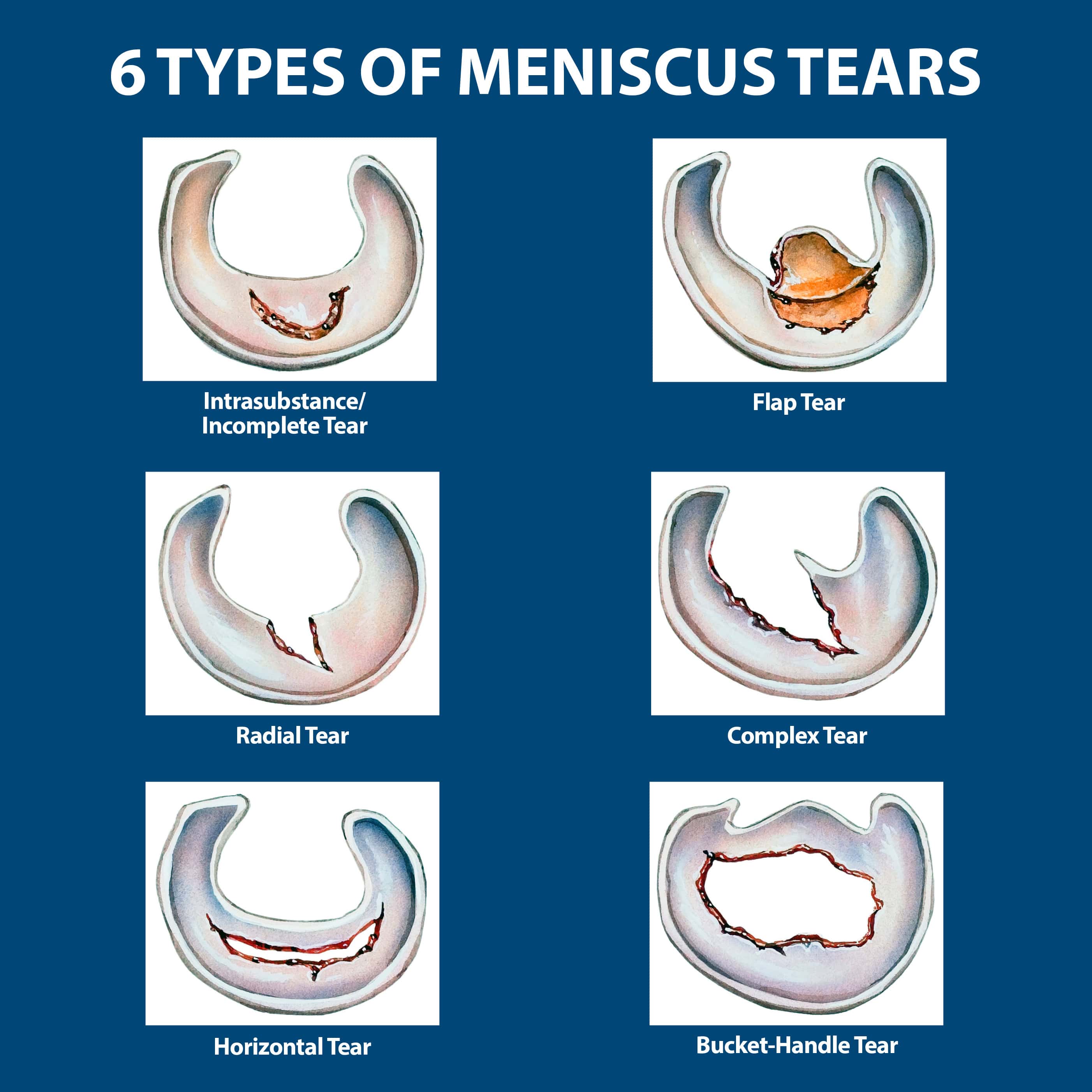
The type of meniscus tear can vary based on its location, shape, and severity. Here are some common types of meniscus tears:
- Horizontal Tear: This type of tear occurs across the width of the meniscus, often resulting from a twisting injury.
- Vertical Tear: This tear runs vertically along the meniscus, often occurring from a direct impact or a sudden twisting motion.
- Radial Tear: This type of tear originates from the outer edge of the meniscus and extends inwards, resembling a spoke on a wheel. It can be caused by a forceful twisting or bending motion.
- Bucket-Handle Tear: A serious type of tear where a large portion of the meniscus is displaced, resembling a bucket handle. It often occurs due to a forceful twisting or impact injury.
- Flap Tear: This tear involves a portion of the meniscus being torn away from the main body, creating a flap. It is often caused by a forceful impact or twisting injury.
- Degenerative Tear: These tears occur over time due to wear and tear on the meniscus, often in individuals over 40.
Causes of Meniscus Tears
Meniscus tears can occur due to various factors, including:
- Sports Injuries: Activities involving twisting, pivoting, and sudden changes in direction can put stress on the meniscus, increasing the risk of tears. Examples include football, basketball, skiing, and tennis.
- Degenerative Changes: As we age, the meniscus can weaken and become more prone to tears. This is often associated with osteoarthritis, a condition that affects the joints.
- Trauma: A direct impact to the knee, such as a fall or a car accident, can cause a meniscus tear.
Example: A basketball player landing awkwardly after a jump might experience a bucket-handle tear, while a skier twisting their knee during a fall might suffer a flap tear.
Symptoms and Diagnosis of Meniscus Tears

A meniscus tear can cause a range of symptoms, depending on the severity of the tear and the individual’s activity level. Some individuals may experience minimal discomfort, while others may have significant pain and limitations in their daily activities.
A careful physical examination and imaging studies are crucial for diagnosing a meniscus tear and determining the best course of treatment.
Physical Examination
A thorough physical examination can help identify signs of a meniscus tear. The doctor will assess your range of motion, palpate the affected knee for tenderness, and perform specific tests to evaluate the stability of your knee joint.
One common test used to diagnose a meniscus tear is the McMurray test. This test involves flexing and extending the knee while rotating the lower leg. A clicking or popping sound, along with pain, may indicate a meniscus tear.
Imaging Studies
Imaging studies, such as magnetic resonance imaging (MRI), play a vital role in confirming the diagnosis and assessing the extent of a meniscus tear.
An MRI provides detailed images of the soft tissues in the knee, including the meniscus. It can reveal the location, size, and severity of the tear.
MRI is considered the gold standard for diagnosing meniscus tears.
The results of the MRI can help guide treatment decisions, such as whether surgery is necessary or if conservative measures, such as rest, ice, compression, and elevation (RICE), are sufficient.
